-
 Mail us
info@myhealthhospitals.com
-
 Toll Free
+91 9111674111
- Book Appointment
info@myhealthhospitals.com
+91 9111674111
Cardiology
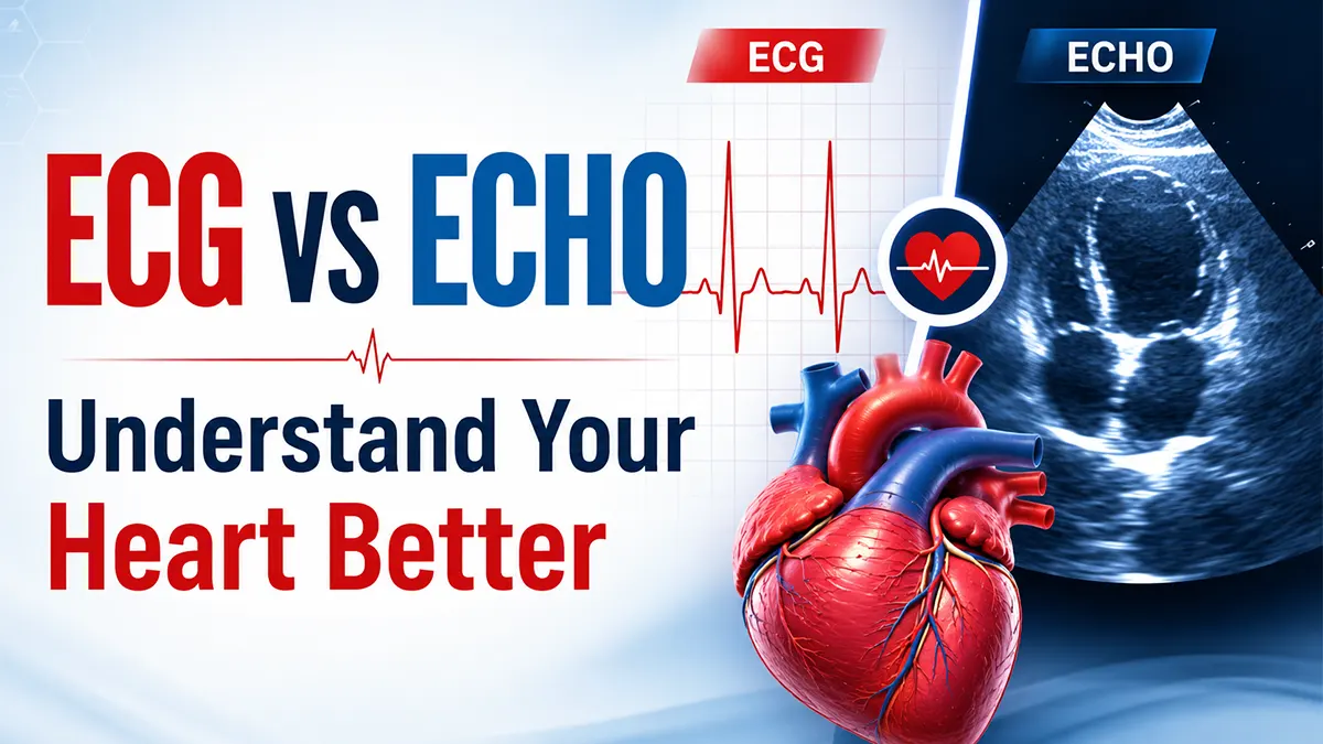
Wondering whether you need an ECG or an ECHO test? While both are essential heart tests, they evaluate different aspects of cardiovascular health. Understanding the difference between an Electrocardiogram (ECG) and an Echocardiogram (ECHO) can help patients make informed decisions and seek timely medical care.
Heart disease continues to be one of the leading causes of hospitalization and premature death worldwide. Conditions such as coronary artery disease, heart failure, arrhythmias, valve disorders, and stroke often develop silently before symptoms become severe. Early diagnosis through advanced cardiac screening plays a critical role in improving treatment outcomes and reducing long-term complications.
Two of the most frequently recommended cardiac investigations are the Electrocardiogram (ECG) and the Echocardiogram (ECHO). Although patients often use these terms interchangeably, these tests serve completely different diagnostic purposes. Each test provides unique information about heart health and helps cardiologists identify specific cardiovascular conditions.
An ECG (Electrocardiogram) measures and records the heart's electrical activity. It helps detect abnormal heart rhythms (arrhythmias), previous or ongoing heart attacks, conduction abnormalities, and other electrical disturbances affecting the heart.
An ECHO (Echocardiogram), on the other hand, uses ultrasound technology to create detailed real-time images of the heart. It allows cardiologists to assess heart chambers, valves, blood flow, heart muscle function, pumping efficiency, and structural abnormalities that cannot be identified through an ECG alone.
Because ECG and ECHO evaluate different aspects of heart function, doctors often recommend both tests together for a comprehensive cardiac assessment. These investigations are commonly advised for patients experiencing chest pain, palpitations, breathlessness, dizziness, high blood pressure, unexplained fatigue, or a family history of heart disease.
At My Health Hospitals, Hyderabad, our experienced cardiology specialists provide advanced ECG testing, 2D Echocardiography, Doppler studies, and preventive heart screening services using modern diagnostic technology. Early cardiac evaluation can help identify heart problems before they become life-threatening and support better long-term cardiovascular health.
Both ECG and Echocardiography are important heart tests, but they evaluate different aspects of cardiac health. Understanding the difference helps patients know why their cardiologist may recommend one or both tests during a heart evaluation.
| Feature | ECG (Electrocardiogram) | ECHO (Echocardiogram) |
|---|---|---|
| Purpose | Measures electrical activity of the heart | Creates images of the heart using ultrasound |
| Duration | 5–10 minutes | 15–30 minutes |
| Pain | Painless | Painless |
| Radiation | No radiation exposure | No radiation exposure |
| Detects | Arrhythmias, heart attacks, conduction abnormalities | Valve disease, heart failure, structural abnormalities |
| Technology | Skin electrodes | Ultrasound waves |
| Output | Electrical waveform tracing | Real-time moving heart images |
An Electrocardiogram (ECG) is a quick, painless heart test that records the electrical activity of the heart. Doctors use an ECG to detect abnormal heart rhythms, heart attacks, conduction disorders, and other cardiac conditions within minutes.
An ECG is one of the most commonly performed cardiac diagnostic tests worldwide because it provides valuable information about how the heart's electrical system is functioning. The test helps cardiologists identify heart problems early, often before symptoms become severe.
Whether performed during a routine health checkup, emergency evaluation, pre-surgery assessment, or heart disease screening, an ECG plays an important role in diagnosing cardiovascular conditions and guiding treatment decisions.
During an ECG, small adhesive electrodes are placed on the chest, arms, and legs. These sensors record the heart's electrical impulses and generate a tracing that allows doctors to evaluate heart rhythm, heart rate, and electrical conduction patterns.
An Echocardiogram (ECHO) is a non-invasive ultrasound test that creates real-time images of the heart. Doctors use an ECHO to evaluate heart valves, heart chambers, blood flow, pumping function, and structural heart abnormalities.
Unlike an ECG, which records the heart's electrical activity, an Echocardiogram provides a visual assessment of how the heart is physically functioning. It helps cardiologists diagnose heart valve disease, heart failure, congenital heart defects, cardiomyopathy, and other structural cardiac conditions.
Because Echocardiography uses harmless ultrasound waves, the test is completely safe, painless, and free from radiation exposure. It is one of the most commonly recommended heart imaging tests for patients experiencing chest discomfort, breathlessness, heart murmurs, swelling in the legs, or abnormal ECG findings.
During an Echocardiogram, a healthcare professional places a handheld device called a transducer on the chest. The transducer sends ultrasound waves that create detailed moving images of the heart on a monitor. Most ECHO tests take between 15 and 30 minutes to complete.
The main difference between ECG and ECHO is that an ECG measures the heart's electrical activity, while an ECHO creates ultrasound images of the heart's structure and function. Both tests are important for diagnosing heart disease, but they evaluate different aspects of cardiovascular health.
If you are wondering whether you need an ECG or an Echocardiogram, the answer depends on your symptoms and your doctor's assessment. ECG is primarily used to identify rhythm and electrical abnormalities, while ECHO helps visualize heart chambers, valves, blood flow, and pumping efficiency.
An ECG records electrical signals generated by the heart during each heartbeat. It is commonly used during emergency evaluations, routine heart screenings, and investigations of chest pain or palpitations.
An Echocardiogram uses ultrasound technology to create detailed real-time images of the heart. It helps cardiologists assess heart structure, valve function, blood flow, and pumping performance.
| Feature | ECG | ECHO |
|---|---|---|
| Purpose | Measures electrical activity | Creates heart images using ultrasound |
| Detects | Arrhythmias, Heart Attacks | Valve Disease, Heart Failure |
| Procedure Time | 5–10 Minutes | 15–30 Minutes |
| Pain | Painless | Painless |
| Radiation | No | No |
| Output | Electrical Wave Tracing | Moving Heart Images |
Doctors recommend an ECG or Echocardiogram when patients experience symptoms such as chest pain, palpitations, breathlessness, dizziness, fainting, or other signs of heart disease. While an ECG evaluates the heart's electrical activity, an ECHO assesses heart structure, valves, blood flow, and pumping function.
The choice between ECG and ECHO depends on the patient's symptoms, medical history, physical examination findings, and cardiovascular risk factors. In many cases, cardiologists recommend both tests together to obtain a complete picture of heart health.
An ECG is usually the first heart test performed when doctors suspect an abnormal heart rhythm, electrical conduction problem, or heart attack.
ECG testing is widely used in emergency departments, outpatient clinics, preventive heart checkups, and cardiovascular risk assessments.
An Echocardiogram is recommended when doctors need detailed information about heart structure, valve function, blood flow, and pumping efficiency.
An ECHO provides detailed structural information that cannot be obtained from an ECG alone and is often used to confirm or further investigate abnormal cardiac findings.

MBBS, MD General Medicine, DM Cardiology
Cardiologist & Interventional Cardiologist
9+ Years of Experience | 500+ Successful Cardiac Procedures
Dr. Vijay Soorampally specializes in preventive cardiology, coronary artery disease, hypertension management, heart failure treatment, angiography, angioplasty, and advanced cardiac interventions. This article has been medically reviewed for clinical accuracy and patient education purposes.
Experiencing chest pain, palpitations, breathlessness, high blood pressure, or other heart-related symptoms? Consult Dr. Vijay Soorampally for expert cardiac evaluation, ECG, 2D ECHO, preventive heart screening, and advanced cardiology care at My Health Hospitals.
Timings: 4:00 PM – 6:00 PM | Location: My Health Hospitals, Hyderabad
If you are experiencing chest pain, palpitations, shortness of breath, or other cardiac symptoms, consult our specialists for a comprehensive heart evaluation. Learn more about our advanced cardiology services in Hyderabad, including ECG, 2D Echocardiography, Doppler studies, preventive heart screening, and treatment for heart disease.
According to the American Heart Association (AHA) , an ECG is one of the most commonly used tests to evaluate heart rhythm abnormalities and detect signs of heart disease.
Looking for heart screening tests in Hyderabad? Learn the difference between ECG and Echocardiogram (ECHO), when they are recommended, and how they help diagnose heart conditions.
Expert-written medical articles from My Health Hospital
Find the Best Hospital Near You
H.No 15-24-212, MIG-212, Rd Number 1, K P H B Phase 1, Kukatpally, Hyderabad, Telangana 500072
H.No:-12-5-30, WhiteHouse, Moula Ali Rd, South Lalaguda, Tarnaka, Secunderabad, Telangana 500017