
2026 Cardiology Guide
2D Echocardiography Test in Hyderabad: Cost, Procedure, Normal EF & Doppler
2D echocardiography (2D Echo) is a non-invasive ultrasound examination that produces moving images of the heart. It helps clinicians assess heart chambers, valves, heart muscle movement and pumping function.
Quick answer: A 2D echo is different from an ECG. ECG records electrical activity, while echocardiography uses ultrasound to assess the heart's structure and function. Doppler can add information about blood flow.
This guide covers 2D echo meaning, why it is done, procedure, preparation, LVEF, common report terms, Doppler echo, ECG comparison and current cost considerations in Hyderabad.
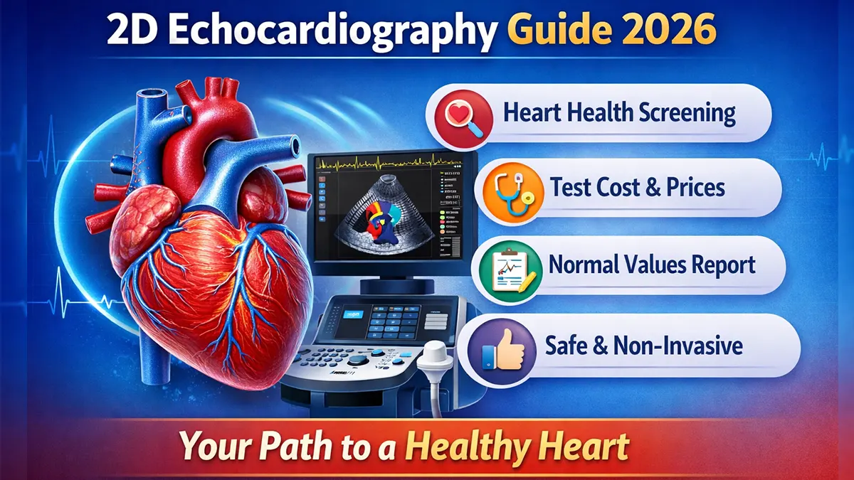
What Is a 2D Echocardiography Test?
It is a specialized heart ultrasound that allows clinicians to assess cardiac anatomy and function in real time.
Heart Chambers
It can assess chamber size, wall movement and overall cardiac structure.
Heart Pumping
Left ventricular ejection fraction (LVEF) and regional wall motion can help assess pumping function.
Valves & Blood Flow
Doppler techniques can assess flow across valves and help identify narrowing or leakage.
Why Is a 2D Echo Test Done?
Doctors may recommend echocardiography when symptoms, examination findings or an existing heart condition require structural and functional assessment.
- Breathlessness, swelling or symptoms that may suggest heart disease
- Heart murmur or suspected valve disease
- Known or suspected cardiomyopathy or heart failure
- Assessment of heart muscle movement or previous cardiac injury
- Congenital heart conditions
- Monitoring a known cardiac condition or response to treatment
- Selected pre-operative or clinical evaluations when indicated
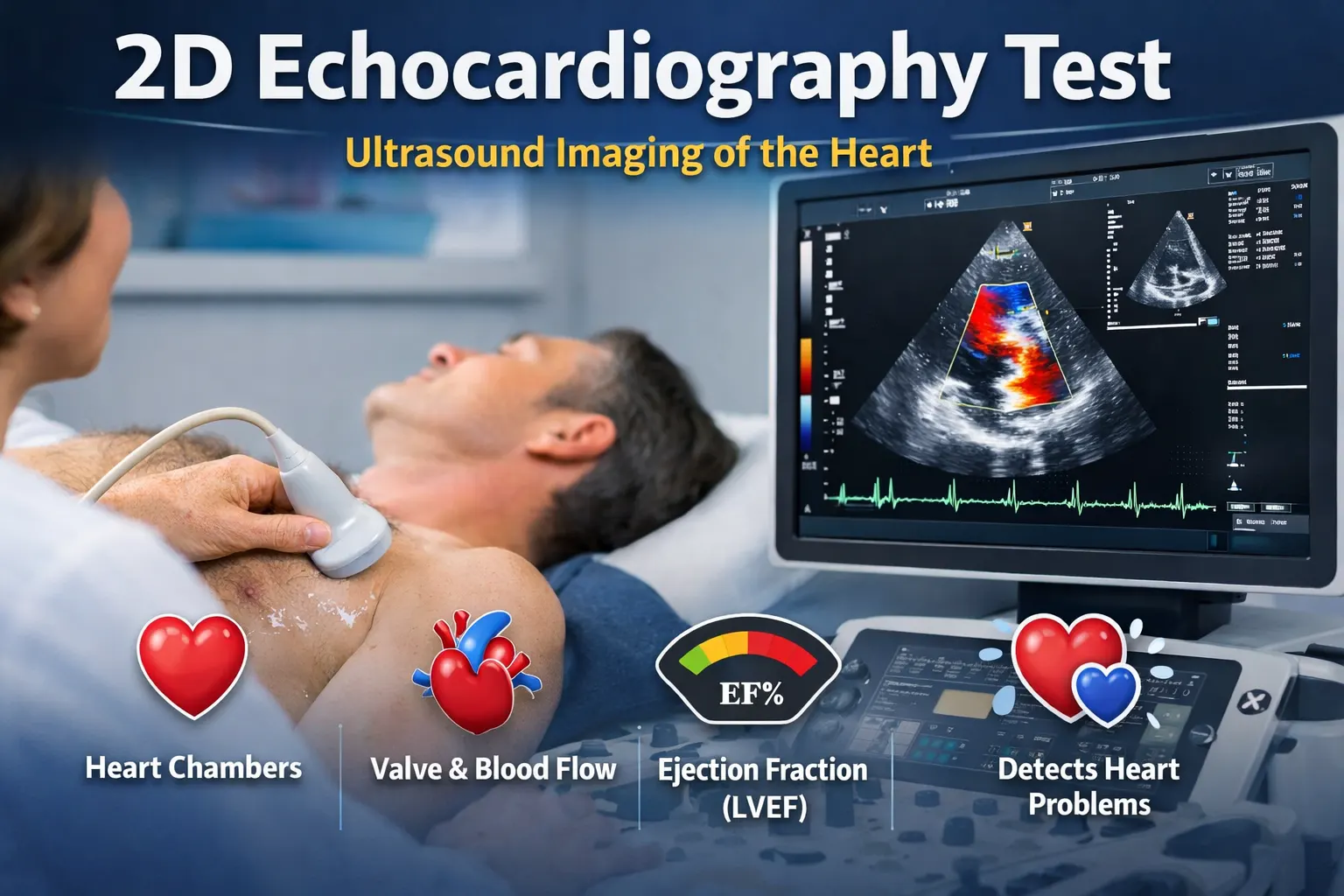
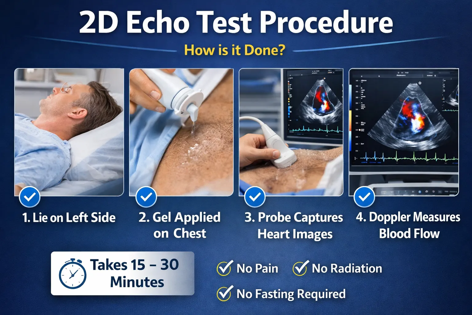
2D Echo Test Procedure: How Is It Done?
A standard transthoracic echocardiogram is usually performed while you are lying down, often with the upper body positioned slightly toward the left.
- Small electrodes may be placed to monitor the heart rhythm during the examination.
- Ultrasound gel is applied to the chest.
- The sonographer or clinician moves the transducer over different areas of the chest.
- Ultrasound waves create moving images of the heart.
- Doppler measurements may be obtained when blood-flow assessment is required.
- The completed images and measurements are reviewed and interpreted by the appropriate clinician.
Preparation: Standard transthoracic echocardiography usually does not require fasting. Follow any specific instructions given by the testing center.
2D Echocardiography With Doppler
Doppler echocardiography adds blood-flow information to standard ultrasound images. It is particularly useful when evaluating valves, abnormal blood-flow patterns and pressure-related findings.
Valve Assessment
Can help assess valve narrowing (stenosis), leakage (regurgitation) and abnormal flow.
Blood-Flow Measurements
Doppler techniques can measure or estimate flow velocities and gradients across selected cardiac structures.
Pressure Estimates
Certain Doppler findings can help estimate pulmonary or intracardiac pressures as part of a complete examination.
Important: Echocardiography is a diagnostic test, not a direct test for coronary artery blockage. The appropriate test depends on the clinical question.
2D Echo Normal Values: Understanding LVEF
One of the most searched 2D echo measurements is left ventricular ejection fraction (LVEF). It estimates the percentage of blood pumped out of the left ventricle with each contraction.
| LVEF | General interpretation |
|---|---|
| 55%–70% | Commonly considered within the normal range. |
| 41%–49% | Mildly reduced range in many clinical frameworks. |
| ≤40% | Reduced ejection fraction; clinical interpretation and cause are important. |
| ≥50% with symptoms | A normal or near-normal EF does not by itself exclude heart disease or heart failure. |
Example: An LVEF of 55% is generally within the normal range. The full report, symptoms and clinical history are more important than one number alone.
Other Report Terms
- Chamber dimensions
- Wall-motion findings
- Valve structure and function
- Diastolic function
Doppler Findings
- Flow velocity
- Valve gradients
- Regurgitation or stenosis
- Estimated pressure findings
What Conditions Can a 2D Echo Help Detect?
Echocardiography can identify or help evaluate many structural and functional heart abnormalities.
- Heart valve disease
- Cardiomyopathy
- Reduced pumping function
- Regional wall-motion abnormalities
- Congenital heart defects
- Pericardial effusion
- Chamber enlargement
- Diastolic dysfunction
- Right-heart abnormalities
- Some pulmonary-pressure findings
- Intracardiac shunts
- Other structural abnormalities
2D Echo Test Cost in Hyderabad (2026)
The price of a 2D echocardiography test varies by diagnostic center, the type of echocardiogram, Doppler requirements, specialist interpretation and whether a specialized study is needed.
Standard 2D Echo
Confirm the current test price directly with the diagnostic center because fees can change.
2D Echo + Doppler
Pricing may differ when Doppler measurements or a more detailed cardiac assessment are included.
Specialized Echo
Fetal or other specialized echocardiographic studies can have different preparation, expertise and pricing requirements.
For Hyderabad: My Health Hospitals has locations serving KPHB/Kukatpally and Tarnaka. Ask the relevant branch about the current test availability, preparation and price before visiting.
How Long Does a 2D Echo Take?
A standard transthoracic echocardiogram commonly takes around 30–60 minutes, depending on the study and image quality required.
Preparation: Usually no fasting for a standard transthoracic echo.
Radiation: Ultrasound-based; no ionizing radiation.
Radiation: Ultrasound-based; no ionizing radiation.
2D Echo vs ECG: What Is the Difference?
| ECG | 2D Echocardiogram |
|---|---|
| Records electrical activity of the heart | Uses ultrasound to show heart structure and movement |
| Helps assess rhythm and electrical patterns | Assesses chambers, valves, pumping function and blood flow |
| Usually very quick | Usually takes longer because detailed images and measurements are obtained |
| Does not directly show heart chambers and valves | Provides structural and functional information |
They are complementary tests. A doctor may recommend one or both depending on symptoms and the suspected condition.
2D Echocardiography: Common Questions
Answers to frequently searched questions about 2D echo tests, EF, Doppler, preparation and reports.
1. What is a 2D echocardiography test?
A 2D echocardiogram is an ultrasound test that creates moving images of the heart and assesses chambers, valves, wall movement and pumping function.
2. What is 2D echo used for?
It helps evaluate symptoms and conditions involving heart structure, valves, heart muscle and pumping function.
3. What is the normal EF in a 2D echo?
An LVEF around 55% to 70% is commonly considered normal, although the complete report and clinical context matter.
4. Is an EF of 55% normal?
Generally yes. An EF of 55% is usually within the normal range.
5. Does 2D echo detect heart blockage?
A resting 2D echo does not directly show coronary blockages. Other tests are used when coronary artery disease needs to be assessed.
6. What is a 2D echo with Doppler?
Doppler adds blood-flow information and helps assess valve flow, gradients and selected pressure-related findings.
7. How long does a 2D echo test take?
A standard study commonly takes around 30 to 60 minutes, depending on the examination.
8. Is 2D echocardiography painful?
It is generally painless. The ultrasound probe is moved over the chest using gel, with some pressure when needed.
9. Is fasting required for a 2D echo?
Usually not for a standard transthoracic echo. Follow the specific instructions given by your center.
10. Is 2D echo safe during pregnancy?
Standard echocardiography uses ultrasound and may be performed during pregnancy when medically indicated.
11. What should I check in a 2D echo report?
Review LVEF, chamber measurements, wall motion, valves, Doppler findings and diastolic function with the interpreting clinician.
12. Can a 2D echo detect heart valve problems?
Yes. It can evaluate valve structure and function and identify many types of narrowing or leakage.
13. Can 2D echo detect heart failure?
It can assess pumping and structural abnormalities that are important in heart-failure evaluation, but diagnosis uses the complete clinical picture.
14. Who may need a 2D echo?
People with relevant symptoms, a heart murmur, suspected or known heart disease, congenital conditions or a need for cardiac monitoring may be referred for an echo.
15. What is the difference between ECG and 2D echo?
ECG records electrical activity; 2D echo uses ultrasound to assess heart structure, movement, valves and pumping function.
16. What is the 2D echo test cost in Hyderabad?
Costs vary by center and test type. Confirm the current price directly with the diagnostic center before booking.
17. Is 2D echo the same as an ultrasound?
A 2D echo is a specialized ultrasound examination designed to evaluate the heart.
18. Can a 2D echo show an enlarged heart?
Yes. Echocardiography can measure heart chambers and assess structural changes.
19. Can a 2D echo detect a hole in the heart?
It can identify many structural defects such as atrial or ventricular septal defects, with Doppler or specialized imaging used when needed.
20. Can 2D echo measure blood pressure inside the heart?
Doppler can estimate certain intracardiac or pulmonary pressures as part of the echocardiographic assessment.
2D Echocardiography in Hyderabad: KPHB & Tarnaka
Patients can contact the appropriate My Health Hospitals branch to confirm current 2D echo availability, pricing, preparation and reporting arrangements.
KPHB / Kukatpally
Convenient for patients in KPHB, Kukatpally and nearby areas who need cardiac diagnostic evaluation or follow-up.
Tarnaka
Patients in Tarnaka and surrounding Hyderabad areas can contact the branch for echocardiography and cardiology-related diagnostic needs.
When to seek urgent medical care: Severe or new chest pain, significant breathing difficulty, fainting, sudden weakness, or other symptoms of a possible heart emergency should be assessed urgently. Do not wait for a routine echocardiogram appointment.